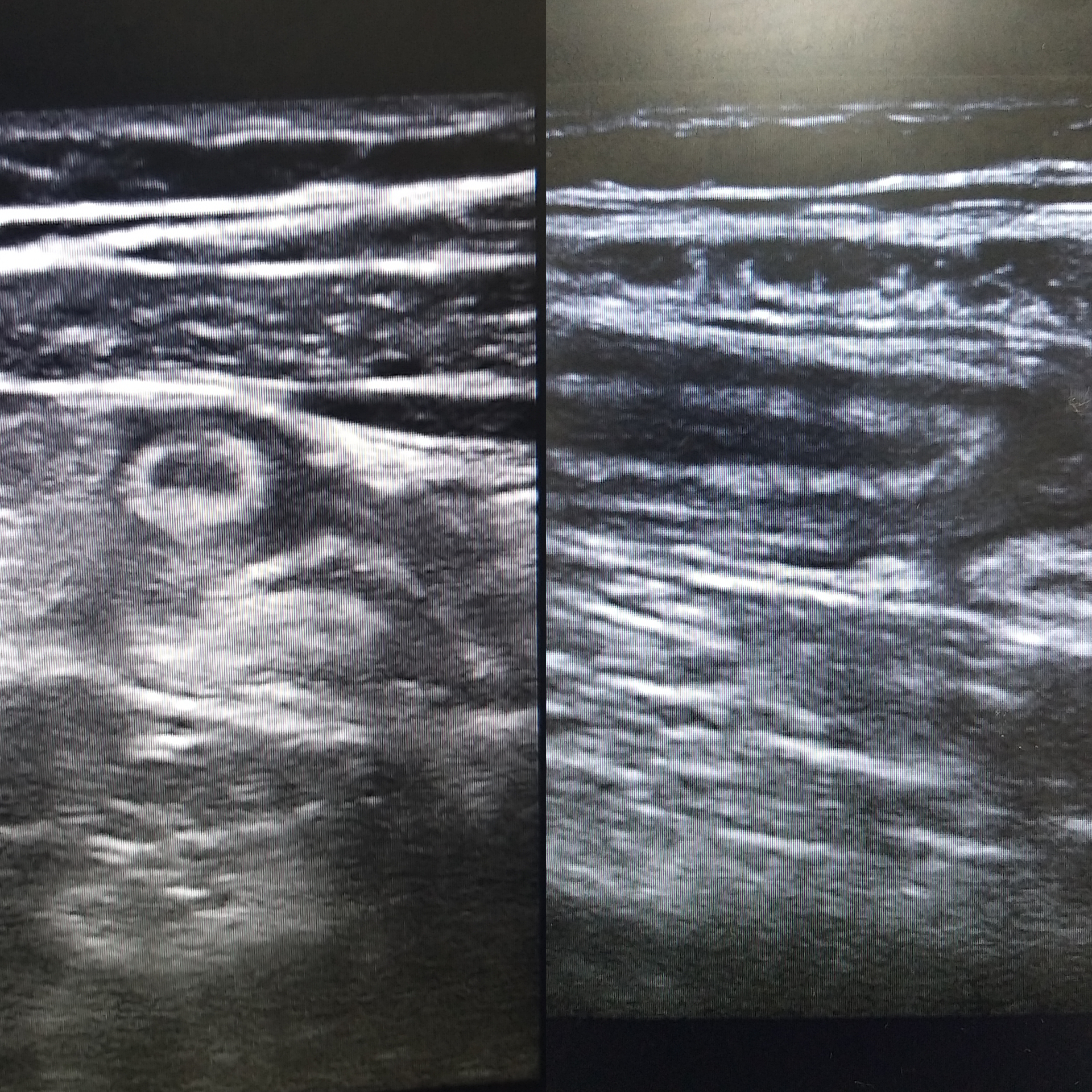
loader image
Close
  • Home
  • About Us
    • Contact Us
  • Gallery
  • Community
    • Resident Corner
      • Blog Posts
      • Hobby Hub
    • Events/Announcements
  • ECREM|Talks
  • FOAMed
  • Exam Resources
    • Exam Prep
    • QOM
  • Research
    • EBM Grand Rounds
    • Clinical Images
    • Resident Publications
  • Frequently Asked Questions (FAQs)
  • Login
  • Register
Menu
  • Home
  • About Us
    • Contact Us
  • Gallery
  • Community
    • Resident Corner
      • Blog Posts
      • Hobby Hub
    • Events/Announcements
  • ECREM|Talks
  • FOAMed
  • Exam Resources
    • Exam Prep
    • QOM
  • Research
    • EBM Grand Rounds
    • Clinical Images
    • Resident Publications
  • Frequently Asked Questions (FAQs)
  • Login
  • Register

Clinical Images

Open for Submissions

Loading...

Buckle Fracture

Name:

Dr. Farnoosh Farzin

A 10 year old female brought to the ED after she sustained a fall on her forearm. On examination, there was swelling and tenderness over the distal forearm. X-ray of the forearm was done and showed torus fracture distal left radial shaft with cortical buckling.

Ingested Key

Name:

Dr. Heba Mustafa Sabri

2 y/o male, came with foreign body ingestion. Abdominal X-ray revealed a metallic density foreign body consistent with ingested key seen overlying the right iliac fossa.

Coin ingestion

Name:

Dr. Farnoosh Farzin

A four year old patient presented to the ED one hour after ingesting 50 fils coin. He was vitally stable. Not in respiratory distress, chest is clear with bilateral air entry, no wheezes or stridor. Child was taken to the OT for esophagogastroduodenoscopy and the coin was successfully removed.

Coffee Bean Sign

Name:

Dr. Farnoosh Farzin

37 year old male, presented to the ED complaining of abdominal pain and nausea for one day. He was vitally stable. X ray abdomen done and showed coffee bean sign suggestive of sigmoid volvulus, and air fluid level.

Spinal Cord Compression

Name:

Dr. Emranur Rahman

26-year-old male Presented to the Emergency Department, post Motor-Vehicle- Collision, with inability to move all his limbs and decreased breath sounds bilaterally. Chest X-ray showed no obvious pneumo- or hemothorax. CT-trauma revealed cervical spine dislocation. Further assessment with MRI revealed spinal cord compression.

Diaphragmatic Hernia

Name:

Dr. Emranur Rahman

63-year-old male presented to ED with worsening chronic dyspnea. Chest X ray done, showed a large left diaphragmatic hernia. On further history patient reported sustaining a road-traffic-accident in the distant past but wasn't diagnosed with any injuries that time despite undergoing extensive imaging

1 2

Submit Your Clinical Image With ECREM

Note: “Please ensure all images submitted conform to appropriate consent, privacy and confidentiality requirements as per your institution. No identifiers, sensitive information or inappropriate content should be included. Acceptance of submission is subject to these conditions and may involve additional questions regarding the content from a member of the ECREM team”.

All published clinical images are peer-reviewed and constitute a scholarly activity according under the Accreditation Council for Graduate Medical Education (ACGME) guidelines.

Our vision follows the slogan بالعلم نرقى , which translates to ‘With knowledge we ascend’. We believe that the sharing and dissemination of knowledge among peers and to others provides an environment where everyone receives the most benefit and through which the overall growth of Emergency Medicine in the community is possible.

Quick Links

  • EBM Grand Rounds
  • Clinical Images
  • Resident Publications
Menu
  • EBM Grand Rounds
  • Clinical Images
  • Resident Publications
  • ECREM|Talks
  • FOAMed
  • Resident Corner
Menu
  • ECREM|Talks
  • FOAMed
  • Resident Corner

Social Links

Instagram Twitter Youtube

ECREM 2021 ©. All right reserved